Gill respiration of animals. Gill breathing is breathing through specialized formations with a dense network of blood vessels. Class Amphibians \u003d Amphibians
Amphibians have two kinds of respiratory organs (not counting the skin): the gills and the lungs. Weakening of gill respiration and the appearance of pulmonary respiration on the scene is already observed in Dipnoi; changes in this direction are observed in Polypterus and Lepidosteus. In amphibians, gill respiration is preserved primarily in the larvae, and then in those Urodela that spend their entire life in water (Perennibranchiata in the former systems). The gill slits are inherited by amphibians from fish-like ancestors. The branchial arches are found in stegocephalus, in larvae, and in some adults (Branchiosauridae). All modern amphibians in the larval state breathe with gills. Normally, they have 5 visceral sacs and the 6th underdeveloped one. But not all of them open outward: there are 4 or even fewer branchial slits. Sometimes the gaps are much smaller than the arcs. The presence of cracks and arcs is evidence of the origin of amphibians from fish. Internal gills, homologous to those of fish, are found, however, only in Anura larvae in the form of short outgrowths of integuments on arcs dividing the gill slits. A soft operculum (operculum), growing from the side of the hyoid arch, covers the gill region from the outside. The operculums of the right and left sides merge with each other on the underside, leaving paired holes in some Anura, and one unpaired one on the left side of the body in most.
In the early stages of development, the Anura larvae and all other amphibians have only external gills, which are apparently homologous to the external gills of Polypterini and Dipnoi larvae. In Apoda and Anura, external gills exist only in the larval period, in the early stages of development, while in Urodela, who returned to aquatic life for the second time, they persist throughout their life. Hence the name for these amphibians is constantly gill (Perennibranchiata), although this name, as has been said, embraces groups of amphibians of various origins. The outer gills are probably inherited by amphibians from cross-finned fish.
Light amphibians have the form of long cylindrical sacks with thin walls (Urodela) or shorter ones (Anura). In legless people, the right lung is much more developed than the left. Lungs appeared in the ancestors of tetrapods long before they went on land. We see the same lungs in lungs. They appeared, obviously, as an additional respiratory organ due to insufficient development of gill respiration, on the one hand, and possibly unfavorable conditions for breathing in dry and rotten waters, on the other. The posterior part of the branchial cavity has developed into an additional respiratory organ. Initially, this organ, which had the appearance of a two-lobed sac that opened on the lower side of the pharynx, was imperfect: its walls had to be swampy, although abundantly supplied with blood, with poorly developed or almost undeveloped septa. Like all branchial protrusions (slits), it had smooth visceral muscles and was innervated by the vagus first.
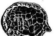
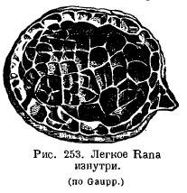
The lungs of amphibians have changed little in comparison: in the aquatic Urodela, the lungs rather act as a hydrostatic apparatus and have a smooth inner surface; the height of their organization is even lower than in Dipnoi. Normally, in amphibians, the inner surface of the lungs is cellular due to the fact that a system of crossbars protrudes into the lung cavity (Fig. 253). It is very interesting that the more terrestrial a particular species is, the more developed are the beams in the lungs: in a toad, the lung is more cellular than frogs. In the genus Ascaphus, living in mountain streams, in water rich in oxygen, cutaneous respiration is highly developed, while the lungs, on the contrary, are small and poorly supplied with blood. A number of amphibians from the suborder Salamandroidea (Salamandrina, Plethodon, Spelerpes, Batrachoseps, etc.) have completely lost their lungs, in return for which pharyngeal and cutaneous respiration has greatly developed. ...
In the simplest case, the lung sacs are interconnected in front, opening directly into the pharynx with a longitudinal slit supported on the sides by cartilaginous strips. These cartilaginous strips, with the help of the muscles attached to them, can expand and narrow the laryngeal gap.
These cartilages originate from the last branchial arch and are found in their simplest form in some Urodela. Cartilages called cricoid cartilages can separate from these cartilages. They can be compared with the arytenoid cartilages (cartilagines arythenoidea) of higher vertebrates. Some Urodela as well as Apoda have a rather long windpipe supported by cartilaginous rings. In Anura, the mucous membrane in the larynx forms the vocal cords. The larynx has complex muscles. At the bottom or at the corners of the mouth are resonators that inflate when croaking.
The respiration mechanism of terrestrial amphibians is rooted in the reflexes observed in fish and aquatic amphibians. The closest thing to the respiration of fish is the respiration of Anura larvae, which have internal gills, an opercular fold and a branchial cavity formed by their fusion, which opens outward with one hole. In addition, in amphibian larvae, the oral cavity is abundantly supplied with blood. By taking water into the mouth and pushing it by raising the jaws through the nostrils, the larvae increase gas exchange in the oral cavity. When the larvae grow up, they rise to the surface, where they swallow air like a ceratode, and by raising the bottom of the oropharyngeal cavity, they push the air into the lungs. In aquatic Urodela, a similar act is observed. When the bottom of the oropharyngeal cavity is lowered and the gill openings are closed behind, water is sucked into the oral cavity through the mouth or nostrils, or through both together. By subsequently raising the floor of the mouth with the nostrils closed, the water is pushed out through the gill slits. Thanks to these movements, the mucous membrane of the mouth and pharynx comes into contact with new masses of water, and the gills get a movement that renews the respiratory environment.
In terrestrial amphibians, the respiratory mechanism is the act of swallowing air due to the lowering of the muscular floor of the oral cavity and pushing it into the lungs due to the raising of the floor. Thus, the respiration of terrestrial amphibians is an act carried out according to the type of a pressure pump, prevailing in lower fish. The immediate basis on which it develops is the respiration mechanism of perpetual-gill amphibians. This latter, observed, for example, in Necturus, must have developed in the distant fish-like ancestors of amphibians. It has already developed a more complex type of terrestrial breathing - Anura.
In lungless salamanders, gas exchange in the intraoral and pharyngeal cavity is highly developed, which occurs with the help of frequent, up to 120-170 oscillations per minute of the diaphragm of the mouth (frogs have 30 of them).
In general, it should be said that pulmonary respiration in amphibians as a whole is an auxiliary mode of respiration. This also contains an indication of its phylogenetic origin.
The respiration of modern amphibians could in no way be the source of the development of respiration of higher Tetrapoda (respiration by raising the ribs, expanding the chest and thus sucking air). The latter type could have arisen, in any case, be outlined in the oldest extinct amphibians, which had long ribs.
Decrease in the number of gills.
An increase in the respiratory surface due to the formation of branchial lobes.
The formation of branchial capillaries.
In the lancelet, the lateral walls of the pharynx are penetrated by numerous (up to 150 pairs) obliquely located branchial slits. The gill arteries approach the intergill septa, and the outgoing gill arteries branch off. When water washes between the gill septa, gas exchange occurs between the passing water and the blood that flows through the thin vessels of the septum. The branchial arteries do not branch into capillaries. In addition, oxygen is released into the animal's body through the capillaries of the skin.
In primary-water vertebrates (jawless and fish), as in the lower chordates, gill slits are formed that connect the pharyngeal cavity with the external environment. In cyclostomes, from the endoderm lining the gill slits, gill sacs are formed (in fish, gills develop from the ectoderm). The inner surface of the sacs is covered with numerous folds - gill petals, in the walls of which a dense network of capillaries branches. The sac opens into the pharynx with an internal narrow channel (in adult lampreys, into the respiratory tube), and with an external one, on the lateral surface of the animal's body. In myxins, there are from 5 to 16 pairs of branchial sacs, in the Bdellostomaceae family, each of them opens outward with an independent opening, and in the myxine family, all external gill ducts on each side merge into one canal, which opens outward with one opening located far behind. Lampreys have 7 pairs of gill sacs, each of which opens outward with an independent opening. Breathing is carried out by rhythmic contractions and relaxation of the muscular wall of the branchial region. In non-feeding lampreys, water enters the respiratory tube from the oral cavity, then washes the petals of the gill sacs, providing gas exchange, and is removed through the external gill ducts. In feeding cyclostomes, water enters and is removed through the external openings of the gill sacs.
The respiratory system of fish has specialized organs of gas exchange - ectodermal gills, which are either located on the intergill septa, as in cartilaginous fish, or directly depart from the gill arches, as in bony fish. The exchange of gases in the gills of vertebrates is built according to the type of "countercurrent systems": when moving on the opposite side, the blood comes into contact with oxygen-rich water, which ensures its effective saturation. The increase in the surface of oxygen absorption due to the formation of gills was accompanied by a decrease in the number of branchial slits in vertebrates as compared with the lower chordates. In whole-headed fish (from cartilaginous fish), a reduction of the intergill septa is observed and a leathery gill cover is formed, covering the outside of the gills. In bony fish, a bony skeleton appears in the operculum, and the intergill septa are reduced, which contributes to more intensive washing of the gill lobes with water. Along with gas exchange, the gills of fish participate in water and salt metabolism, in the elimination of ammonia and urea from the body. The skin, swim bladder, supraopharyngeal labyrinths, and specialized sections of the intestinal tube function as additional respiratory organs in certain groups of fish. In lung-breathing and multi-feather-like fish, organs of air respiration appear - the lungs. The lungs arise as paired outgrowths of the abdominal part of the pharynx in the region of the last branchial slit and are connected with the esophagus by a short canal. The walls of this outgrowth are thin and abundantly supplied with blood.
Trends in the evolution of pulmonary respiration
The emergence and differentiation of the airways.
Lung differentiation and increase in respiratory surface.
The development of auxiliary organs (chest).
In amphibians, the following are involved in the absorption of oxygen and the release of carbon dioxide: in the larvae - the skin, external and internal gills, in adults - the lungs, skin and mucous membrane of the oropharyngeal cavity. In some species of tailed amphibians (sirens, proteas) and in adults, gills are preserved and the lungs are underdeveloped or reduced. The ratio of pulmonary and other types of gas exchange is not the same: in species of humid habitats, skin respiration dominates in gas exchange; in inhabitants of dry places, most of the oxygen enters through the lungs, but the skin plays an essential role in the release of carbon dioxide. The respiratory system of adult amphibians includes the oropharyngeal, laryngeal-tracheal cavities and saccular lungs, the walls of which are braided with a dense network of capillaries. In tailless amphibians there is a common laryngeal-tracheal chamber; in caudates, it is divided into the larynx and trachea. Arytenoid cartilages appear in the larynx, which support its wall and vocal cords. The lungs of tailed amphibians are two thin-walled sacs without partitions. In tailless, inside the lung sacs, there are partitions on the walls that increase the surface of gas exchange (cellular lungs). Amphibians have no ribs, and the act of breathing occurs by forcing air during inhalation (by increasing and then decreasing the volume of the oropharyngeal cavity) and pushing out air during exhalation (due to the elasticity of the walls of the lungs and abdominal muscles).
In reptiles, further differentiation of the airways and a significant increase in the functional surface of gas exchange in the lungs are noted. The airways are divided into the nasal cavity (it is combined with the oral cavity, but in crocodiles and turtles, these cavities are separated by the bony palate), the larynx, the trachea and two bronchi. The walls of the larynx are supported by paired arytenoid and unpaired cricoid cartilages. In lizards and snakes, the inner walls of the pulmonary sacs have a folded cellular structure. In turtles and crocodiles, a complex system of septa protrudes into the inner cavity of the lung so deeply that the lung acquires a spongy structure. The ribcage is formed: the ribs are movably connected to the spine and sternum, the intercostal muscles develop. The act of breathing is carried out due to a change in the volume of the chest (costal breathing). Turtles retain the oropharyngeal type of air injection. In aquatic turtles in water, additional respiratory organs are the outgrowths of the pharynx and cloaca (anal vesicles) rich in capillaries. Reptiles lack skin respiration.
In birds, the airways are represented by the nasal cavity, the larynx, which is supported by arytenoid and cricoid cartilages, a long trachea and the bronchial system. The lungs are small, dense and not very extensible and are accreted to the ribs on the sides of the spinal column. Primary bronchi are formed when the lower part of the trachea is divided and enter the tissue of the corresponding lung, where they break up into 15–20 secondary bronchi, most of which end blindly, and some of them communicate with air sacs. Secondary bronchi are interconnected by smaller parabronchuses, from which many thin-walled cellular bronchioles depart. The bronchioles braided by blood vessels form the morphofunctional structure of the lung. Air sacs are associated with the lungs of birds - transparent elastic thin-walled outgrowths of the mucous membrane of the secondary bronchi. The volume of the air sacs is about 10 times the volume of the lungs. They play a very important role in the implementation of a kind of breathing act of birds: air with a high oxygen content enters the lungs both during inhalation and exhalation - "double breathing". In addition to intensifying breathing, air bags prevent the body from overheating during intense movement. The increase in intra-abdominal pressure during expiration promotes defecation. Diving birds, by increasing the pressure in the air sacs, can decrease the volume and thereby increase the density, which facilitates immersion in water. Cutaneous respiration in birds is absent.
In mammals, further differentiation of the airways is observed. The nasal cavity, the nasopharynx are formed, the entrance to the larynx is covered by the epiglottis (in all terrestrial vertebrates, except for mammals, the laryngeal slit is closed by special muscles), thyroid cartilage appears in the larynx, then the trachea, which branches into two bronchi, going into the right and left lungs. In the lungs, the bronchi repeatedly branch and end with bronchioles and alveoli (the number of alveoli is from 6 to 500 million), this significantly increases the respiratory surface. Gas exchange occurs in the alveolar passages and alveoli, the walls of which are densely braided with blood vessels. The morphofunctional unit of the mammalian lung is the pulmonary acinus, which is formed as a result of branching of the terminal bronchiole. The rib cage is formed, which is separated from the abdominal cavity by the diaphragm. The number of respiratory movements from 8 to 200. Respiratory movements are carried out in two ways: by changing the volume of the chest (costal breathing) and due to the activity of the diaphragmatic muscle (diaphragmatic breathing). In higher mammals, cutaneous respiration is developed through the system of cutaneous capillaries, which plays an important role in gas exchange.
The set of processes that ensure the consumption of O 2 and the release of CO 2 in the body is called breathing... There are processes of external and internal respiration. External respiration provides the exchange of gases between the body and the external environment, internal respiration - the consumption of O2 and the release of CO 2 by the cells of the body.
The factor that ensures the diffusion of gases through the respiratory surfaces is the difference in their concentrations. The movement of dissolved gases occurs in the direction from the area with their high concentration to the area of \u200b\u200blow concentration.
In small organisms, gas exchange, as a rule, is carried out diffusely over the entire surface of the body (or cell). In larger animals, gases are transported to the tissues either directly (tracheal system of insects) or using special vehicles (blood, hemolymph).
The amount of oxygen entering the tissues of an animal depends on the area of \u200b\u200bthe respiratory surface and the difference in oxygen concentration on them. Therefore, in all respiratory organs, the growth of the respiratory epithelium is observed. To maintain a high gradient of oxygen diffusion on the exchange membrane, it is necessary to move the medium (ventilation). It is provided by respiratory rhythmic movements of the whole body of the animal (small-bristled tubule worm, leeches) or certain parts of it (crustaceans), as well as the work of the ciliary epithelium (mollusks, lancelet).
A number of fairly large animals do not have specialized respiratory organs. Their gas exchange is carried out through moist skin, supplied with an abundant network of blood vessels (earthworm). Cutaneous respiration as an additional one is characteristic of animals with specialized respiratory organs. For example, in eels with gills, 60% of the oxygen demand is provided by cutaneous respiration, in frogs with lungs, this value is more than 50%.
The respiratory organs in the aquatic environment are the gills, in the ground-air environment - the lungs and trachea.
Gills are organs located outside the body cavity in the form of epithelial surfaces permeated with a dense network of blood capillaries. Gill respiration is characteristic of polychaete annelids, most molluscs, crustaceans, fish, and amphibian larvae. The gill respiration is most effective in fish. It is based on counterflow phenomenon: the blood in the capillaries of the gill lobes flows in the opposite direction to the flow of the ox that washes the gills.
Lungsare usually internal organs and protected from drying out. There are two types of them: diffusion and ventilation... In the lungs of the first type, gas exchange is carried out only by diffusion. Relatively small animals have such lungs: pulmonary mollusks, scorpions, spiders. Only terrestrial vertebrates have ventilation lungs.
The complication of the structure of the lungs in the order from amphibians to mammals is associated with an increase in the area of \u200b\u200bthe respiratory epithelium. So, in amphibians, 1 cm 3 of lung tissue has a total gas exchange surface of 20 cm 2. A similar indicator for the epithelium of the human lungs is 300 cm 2.
Simultaneously with the increase in the respiratory surface, there is an improvement in the ventilation mechanism of the lungs, which, starting with reptiles, is carried out by changing the volume of the chest, and in mammals - with the participation of the muscles of the diaphragm. These adaptations made it possible for warm-blooded animals (birds and mammals) to dramatically increase their metabolic rate.
The third type of respiratory system is trachea... They are thin-walled, branching, non-collapsing invaginations into the body, filled with air. The trachea communicate with the external environment through openings in the cuticle - spiracles. Insects most often have 12 pairs: 3 pairs on the chest and 9 pairs on the abdomen. The spiracles can close or open depending on the amount of oxygen. With a high degree of development of the tracheal system (in insects), its numerous branches entwine all internal organs and directly provide gas exchange in tissues. The fundamental difference between tracheal respiration and pulmonary and branchial respiration is that it does not require the participation of blood as a transport mediator in gas exchange.
The tracheal system is able to maintain a sufficiently high level of tissue respiration, thereby ensuring a high physiological activity of the insect.
Ventilation of the trachea in insects in the absence of flight is carried out most often by rhythmic contractions of the abdomen, in flight it is enhanced by movements of the chest.
Aquatic larvae of some insects breathe with tracheal gills... In this case, the tracheal system is devoid of spiracles i.e. it is closed and filled with air. The branches of a closed tracheal system enter the "gills" - appendages with a large surface and a thin cuticle that allows gas exchange between water and air in the tracheal system. Such tracheal gills are, for example, in mayfly larvae. In the larvae of some dragonflies, the tracheal gills are located in the rectal cavity, and the insect ventilates them, drawing water into the intestine and pushing it back.
Respiratory system evolution
The stages of the breathing process
Breath - a set of processes that ensure the supply of oxygen from the environment to the body, which is necessary for the oxidation of organic substances in the mitochondria of the cell, and the release of carbon dioxide
Breathing types:

Breath type:
Cellular.
Organisms: unicellular animals (amoeba, euglena green, infusoria shoe); coelenterates (jellyfish, coral polyps); some worms.
Single-celled organisms absorb oxygen dissolved in water through the entire surface of the body by diffusion.
Oxygen is involved in the breakdown of complex organic substances, as a result of which the energy that is necessary for the life of the animal is released.
The carbon dioxide formed as a result of breathing is also released outside through the entire surface of the body.
Tracheal breathing is breathing with the help of a system of combined tubule-trachea that penetrate the entire body.
Organisms: class Insects (bugs, butterflies, grasshoppers, flies)
The abdomen of the insect is divided into 5–11 parts (segments). On each of them there is a pair of small holes - spiracle. Branching tubes extend from each spiracle inward - tracheathat permeate the entire body of the insect. Watching the May beetle, you can notice how its abdomen decreases in volume, then increases. These are respiratory movements. When inhaling, air containing oxygen enters the body through the spiracles, and when exhaling, air saturated with carbon dioxide comes out.
In spiders (class Arachnids), the respiratory organs are represented not only by the trachea, but also by the pulmonary sacs, which communicate with the external environment through the respiratory openings.
Gill breathing is breathing using specialized formations with a dense network of blood vessels.
Organisms: many aquatic creatures (fish, crayfish, mollusks)
Fish breathe oxygen dissolved in water with the help of special branched skin outgrowths called gills.
Fish constantly swallow water. From the oral cavity, water passes through the gill slots, is washed by the gills and out of the gill covers comes out. Gillsconsist of branchial arches and gill petalsthat are pierced by a multitude of blood vessels. Oxygen enters the blood from the water that washes the gills, and carbon dioxide is removed from the blood into the water. The gills inside the body are called internal gills.
Some animals, such as amphibians, have dense bundles of gills on the surface of the body. Such gills are called - outdoor. Such is the structure of Proteus - a blind cave animal from the western regions of Yugoslavia, and axolotls (which in general appearance are similar to newts) - their homeland is Mexico.
Class Amphibians \u003d Amphibians.
The first terrestrial vertebrates that still retained a connection with the aquatic environment. The class has 3900 species and includes 3 orders: caudate (salamanders, newts), legless (tropical worms) and tailless (toads, tree frogs, frogs, etc.).
Secondary aquatic animals. Since the amniotic cavity is absent in the egg (together with cyclostomes and amphibians, these are anamnias), they multiply in the water, where the initial stages of their development pass. At different stages of the life cycle, amphibians lead a terrestrial or semi-aquatic lifestyle, distributed almost everywhere, mainly in areas with high humidity along the banks of fresh water bodies and on moist soils. Among amphibians there are no forms that could live in saltwater. Various modes of movement are characteristic: species are known that make fairly long jumps, move in steps or “crawl”, devoid of limbs (worms).
The main signs of amphibians.
Amphibians retained many features of their purely aquatic ancestors, but along with this, they acquired a number of features characteristic of a real terrestrial vertebrate.
Tailed and tailless are characterized by larval development with gill breathing in fresh water (frog tadpoles) and their metamorphosis into an adult breathing lungs. In legless hatching, the larva takes the form of an adult animal.
The circulatory system is characterized by two circles of blood circulation. The heart is three-chambered. It has one ventricle and two atria.
Separate the cervical and sacral spine, having one vertebra.
Adult amphibians are characterized by paired limbs with articulated joints. The limbs are five-fingered.
The skull with two occipital condyles is articulated with the cervical vertebra.
The pelvic girdle is tightly attached to the transverse processes of the sacral vertebra.
The eyes have moving eyelids and blinking membranes to protect the eyes from clogging and drying. Due to the convex cornea and flattened lens, accommodation is improved.
The forebrain is enlarged and divided into two hemispheres. The midbrain and cerebellum are underdeveloped. 10 pairs of cranial nerves leave the brain.
The skin is bare, i.e. devoid of any horn or bone formations, permeable to water and gases. Therefore, it is always wet - oxygen first dissolves in the liquid covering the skin, and then diffuses into the blood. The same thing happens with carbon dioxide, but in the opposite direction.
Kidneys, like fish, primary \u003d mesonephric.
To capture the sound waves of the air, a tympanic membrane appears, behind it is the middle ear (tympanic cavity), in which the auditory ossicle is located - the stirrup, which conducts vibrations to the inner ear. The Eustachian tube communicates with the middle ear cavity. Hoans appear - the internal nostrils, the nasal passages become through.
The body temperature is unstable (poikilothermy) depends on the ambient temperature and only slightly exceeds the latter.
Aromorphoses:
Lungs and pulmonary breathing appeared.
The circulatory system has become more complex, the pulmonary circle of blood circulation has developed, i.e. amphibians have two circles of blood circulation - large and small. The heart is three-chambered.
Formed paired five-fingered limbs, which are a system of levers with articulated joints and designed to move on land.
The cervical spine was formed, providing movement of the head, and the sacral region - the place of attachment of the pelvic girdle.
The middle ear, eyelids, and choans appeared.
Muscle differentiation.
Progressive development of the nervous system.
Phylogeny.
Amphibians descended from the ancient brush-headed fish in the Devonian period of the Paleozoic era about 350 million years ago. The first amphibians - ichthyostegs - in appearance resembled modern caudate amphibians. In their structure there were features characteristic of fish, including rudiments of the gill cover and organs of the lateral line.
Cover. Two-layer. The epidermis is multilayered, the corium is thin, but abundantly equipped with capillaries. Amphibians retained the ability to produce mucus, but not by individual cells, like most fish, but by the mucous glands of the alveolar type. In addition, amphibians often have granular glands with toxic secretions of varying degrees of toxicity. The skin color of amphibians depends on special cells - chromatophores. These include melanophores, lipophores and iridocytes.
Under the skin of the frogs, there are extensive lymphatic gaps - reservoirs filled with tissue fluid and allowing, under adverse conditions, to accumulate a supply of water.
Skeleton It is divided into axial and incremental, as in all vertebrates. The vertebral column is more differentiated into departments than in fish and consists of four departments: cervical, trunk, sacral and caudal. The cervical and sacral sections have one vertebra each. The tail vertebrae of the tailless vertebrae are usually seven, and all the caudal vertebrae (approximately 12) merge into a single ossicle - urostyle. In caudate 13 - 62 trunk and 22 - 36 caudal vertebrae; in legless, the total number of vertebrae reaches 200 - 300. The presence of a cervical vertebra is important, because unlike fish, amphibians cannot turn their bodies around so quickly, and the cervical vertebra makes the head moveable, but with a small amplitude. Amphibians cannot turn their heads, but they can tilt them.
Vertebrae in different amphibians can vary in type. In legless and lower caudate vertebrae are amphibian, with a preserved chord, like in fish. In higher caudate vertebrae, opistocellular, i.e. the front of the body is curved, and the back is concave. In tailless, on the contrary, the front surface of the vertebral bodies is concave, and the back is curved. Such vertebrae are called whole. The presence of articular surfaces and articular processes provides not only a strong connection of the vertebrae, but also makes the axial skeleton movable, which is important for the movement of caudate amphibians in water without the participation of limbs, due to lateral bends of the body. In addition, vertical movements are possible.
The amphibian skull is, as it were, a modified skull of bony fish, adapted to terrestrial existence. The brain skull is retained predominantly by cartilage for life. The occipital region of the skull contains only two lateral occipital bones, which are carried along the articular condyle, with which the skull is attached to the vertebrae. The visceral amphibian skull undergoes the greatest transformations: secondary upper jaws appear; formed by the maxillary (maxillary) and maxillary bones. Reduction of gill respiration led to a radical change in the hyoid arch. The sublingual arch is transformed into an element of the hearing aid and the hyoid plate. Unlike fish, the amphibian visceral skull directly grows with the sky-square cartilage to the bottom of the brain skull. This type of direct connection of the components of the skull without the participation of the elements of the hyoid arch is called autostyly. Elements of the gill cover in amphibians are absent.
The additional skeleton includes bones of the belts and free limbs. Like fish, the bones of the amphibian shoulder girdle are located in the thickness of the muscles that bind them to the axial skeleton, but the belt itself is not directly connected to the axial skeleton. The belt provides support for the free limb.
All terrestrial animals constantly have to overcome gravity, which fish do not need to do. The free limb serves as a support, allows you to raise the body above the surface and provides movement. The free limbs consist of three sections: proximal (one bone), intermediate (two bones) and distal (a relatively large number of bones). Representatives of different classes of terrestrial vertebrates have structural features of one or another free limb, but they are all secondary.
In all amphibians, the proximal part of the free forelimb is represented by the humerus, the intermediate by the ulnar and radial bones of the caudate and the unified bone of the forearm (it is formed as a result of fusion of the ulna and radius) in the tailless. The distal section is formed by the wrist, metacarpus and phalanges of the fingers.
The belt of the hind limbs articulates directly with the axial skeleton, with its sacral section. A reliable and rigid connection of the pelvic girdle with the spinal column ensures the functioning of the hind limbs, which are more important for the movement of amphibians.
Muscular system different from the muscle system of fish. The trunk muscles retain a metameric structure only in the legless. In caudate metamerism of segments is disturbed, and in tailless amphibians, sections of muscle segments begin to separate, differentiating into ribbon-like muscles. The mass of muscles of the limbs increases sharply. In fish, the movements of the fins are mainly provided by the muscles located on the body, while the five-fingered limb moves due to the muscles located in it itself. A complex system of muscles appears - antagonists - muscles of the flexors and extensors. Segmented muscles are present only in the spinal column. The muscles of the oral cavity (chewing, tongue, bottom of the oral cavity) are becoming more sophisticated and specialized, not only participating in the capture and swallowing of food, but also providing ventilation of the oral cavity and lungs.
Body cavity - whole. In amphibians, due to the disappearance of the gills, the relative position of the pericardial cavity has changed. She was pushed to the bottom of the chest into the area covered by the sternum (or coracoid). Above it in a pair of coelomic channels are the lungs. Cavities containing the heart and lungs. Pleurocardial membrane separates. The cavity in which the lungs are located communicates with the main coelom.
Nervous system. The brain of the ichthyopsid type, i.e. the main integrating center is the midbrain, but the amphibian brain has a number of progressive changes. The amphibian brain has five sections and differs from the brain of fish, mainly in the large development of the forebrain, the complete separation of its hemispheres. In addition, the nerve substance is already lining apart from the bottom of the lateral ventricles as well as the sides and the roof, forming the cerebral arch - archipallium. The development of archipallium, accompanied by increased ties with the diencephalon and especially the midbrain, leads to the fact that associative activity that regulates behavior is carried out in amphibians not only by the medulla oblongata and midbrain, but also by the forebrain hemispheres. The elongated hemispheres in front have a common olfactory lobe, from which two olfactory nerves originate. Behind the forebrain is the diencephalon. On its roof is the pineal gland. On the underside of the brain there is a cross of optic nerves (chiasm). From the bottom of the diencephalon, a funnel and pituitary gland (lower cerebral gland) departs.
The midbrain is represented as two round visual lobes. An underdeveloped cerebellum lies behind the visual lobes. Immediately behind it is the medulla oblongata with a rhomboid fossa (fourth ventricle). The medulla oblongata gradually passes into the spinal cord.
In amphibians, 10 pairs of brain nerves depart from the brain. The eleventh pair is not developed, and the twelfth departs outside the skull.
Real frog spinal nerves 10 pairs. Three anterior ones participate in the formation of the brachial plexus innervating the forelimbs, and four hind pairs in the formation of the lumbosacral plexus innervating the hind limbs.
Sensory organs provide guidance to amphibians in water and on land.
The organs of the lateral line are found in all larvae and in adults with an aquatic lifestyle. Represented by an accumulation of sensitive cells with nerves suitable for them, which are scattered throughout the body. Sensitive cells perceive temperature, pain, tactile sensations, as well as changes in humidity and chemical composition of the environment.
The organs of smell. Amphibians on each side of the head have a small outer nostril that leads into an elongated sac ending in the inner nostril (choana). Joans open in front of the roof of the oral cavity. In front of the choan left and right there is a bag that opens into the nasal cavity. This is the so-called vomeronasal organ. It has a large number of sensory cells. Its function is to obtain olfactory information about food.
The organs of vision have a structure characteristic of the terrestrial vertebral. This is expressed in the convex shape of the cornea, the crystalline lens in the form of a biconvex lens, in the moving eyelids, which protect the eyes from drying out. But accommodation, as in fish, is achieved by moving the lens through contraction of the ciliary muscle. The muscle is located in the annular roller surrounding the lens, and when it contracts, the frog lens extends somewhat forward.
The hearing organ is arranged on the ground type. The second section appears - the middle ear, in which the auditory ossicle, the first appearing in vertebrates, is placed - the stirrup. The drum cavity is connected to the pharyngeal region by the Eustachian tube.
The behavior of amphibians is very primitive, conditioned reflexes are developed slowly, and quickly fade away. The motor specialization of reflexes is very small, so the frog cannot form a protective reflex of pulling away one leg, and when irritating one limb, it pulls with both legs.
Digestive system begins with a mouth opening leading to the oropharyngeal cavity. It has a muscular tongue. The ducts of the salivary glands open into it. The tongue and salivary glands first appear in amphibians. The glands serve only to wet the food lump and do not participate in the chemical processing of food. On the intermaxillary, maxillary bones, the vomer, simple conical teeth are located, which are attached to the bone with the base. The digestive tube differentiates into the oropharyngeal cavity, a short esophagus, which performs the function of carrying food into the stomach, and a voluminous stomach. Its pyloric part passes into the duodenum - the beginning of the small intestine. In the loop between the stomach and duodenum lies the pancreas. The small intestine smoothly passes into the large intestine, which ends with a pronounced rectum that opens into the cloaca.
The digestive glands are the liver with the gall bladder and the pancreas. The ducts of the liver together with the duct of the gallbladder open into the duodenum. The pancreatic ducts flow into the gallbladder duct, i.e. this gland does not have independent communication with the intestines.
T.O. the amphibian digestive system differs from the similar fish system in the longer digestive tract, the final part of the colon opens into the cloaca.
Circulatory system closed. Two circles of blood circulation. The heart is three-chambered. In addition, there is a venous sinus in the heart that communicates with the right atrium, and an arterial cone leaves on the right side of the ventricle. Three pairs of vessels homologous to the branchial arteries of fish depart from it. Each vessel begins with an independent opening. All three vessels of the left and right sides go first with a common arterial trunk surrounded by a common membrane, and then branch.
The vessels of the first pair (counting from the head), homologous to the vessels of the first pair of gill arteries of fish, are called the carotid arteries, which carry blood to the head. Through the vessels of the second pair (homologous to the second pair of gill arteries of the fish) - the arches of the aorta - the blood goes to the back of the body. Subclavian arteries departing from the arches of the aorta, carrying blood to the forelimbs.
Through the vessels of the third pair, homologous to the fourth pair of gill arteries of fish - the pulmonary arteries - blood is sent to the lungs. A large cutaneous artery departs from each pulmonary artery, through which blood is sent to the skin for oxidation.
Venous blood from the anterior end of the body is collected along two pairs of jugular veins. The latter, merging with the skin veins that have already taken in the subclavian veins, forms two anterior vena cava. They carry mixed blood into the venous sinus, as arterial blood moves through the skin veins.
Amphibians have one blood circulation, their circulatory system is similar to the circulatory system of fish.
Amphibians have a new circulatory organ - the red bone marrow of the tubular bones. Red blood cells are large, nuclear, white blood cells are not the same in appearance. There are lymphocytes.
Lymphatic system. In addition to the lymphatic bags located under the skin, there are lymphatic vessels and hearts. One pair of lymph hearts is placed near the third vertebra, the other near the cloacal foramen. The spleen, which looks like a small round body of red color, is located on the peritoneum near the beginning of the rectum.
Respiratory system. It is fundamentally different from the respiratory system of fish. In adults, the respiratory organs are the lungs and skin. The respiratory tract due to the absence of the cervical region is short. Represented by the nasal and oropharyngeal cavities, as well as the larynx. The larynx opens directly into the lungs with two holes. Due to the reduction of the ribs, the lungs are filled by swallowing air - according to the principle of a discharge pump.
The anatomically respiratory system of amphibians includes the oropharyngeal cavity (upper airways) and the laryngeal - tracheal cavity (lower ways), which directly passes into the saccular lungs. The lung in the process of embryonic development is formed as a blind outgrowth of the anterior (pharyngeal) section of the digestive tube, and therefore remains in the adult state associated with the pharynx.
T.O. the respiratory system in terrestrial vertebrates is anatomically and functionally divided into two sections - the airway system and the respiratory section. The airways provide two-way air transport, but do not participate in the gas exchange itself, the respiratory department carries out gas exchange between the internal environment of the body (blood) and atmospheric air. Gas exchange occurs through the surface fluid and is passive in accordance with the concentration gradient.
The system of gill covers becomes unnecessary, therefore, the gill apparatus in all terrestrial animals is partially modified, its skeletal structures are partly part of the skeleton (cartilage) of the larynx. Ventilation of the lungs is carried out due to forced movements of special somatic muscles during the respiratory act.
Excretory system as in fish, it is represented by primary, or trunk kidneys. These are compact bodies of a reddish-brown color, lying on the sides of the spine, and not ribbon-like, like in fish. From each kidney a thin wolf channel extends to the cesspool. In female frogs, it serves only as the ureter, while in males it serves as the ureter and vas deferens. In the cesspool, the Wolf channels are opened by independent openings. Also separately opens in the cesspool and the bladder. The final product of nitrogen metabolism in amphibians is urea. In aquatic amphibian larvae, the main product of nitrogen metabolism is ammonia, which in the form of a solution is excreted through the gills and skin.
Amphibians are hyperosmotic animals with respect to fresh water. As a result, water constantly enters the body through the skin, which does not have mechanisms to prevent this, as in other terrestrial vertebrates. Sea water is hyperosmotic in relation to the osmotic pressure in the tissues of amphibians, when placed in such an environment, water through the skin will leave the body. That is why amphibians cannot live in sea water and die in it from dehydration.
The reproductive system. In males, the reproductive organs are represented by a pair of rounded whitish testes adjacent to the abdominal surface of the kidneys. Thin vas deferens the tubules from the testes to the kidneys. Sex products from the testis through these tubules are sent to the bodies of the kidneys, then to the Wolf channels and through them to the cesspool. Before flowing into the cesspool, the wolf channels form a small expansion - seminal vesicles, which serve for temporary deposition of sperm.
The reproductive organs of females are represented by paired ovaries of a granular structure. Above them are fat bodies. They accumulate nutrients that ensure the formation of reproductive products during hibernation. In the lateral parts of the body cavity are strongly convoluted light oviducts, or Müller channels. Each oviduct into the body cavity in the heart region opens with a funnel; the lower uterine part of the oviducts is sharply expanded and opens into the cloaca. The ripened eggs fall through the rupture of the walls of the ovary into the body cavity, then are captured by the funnels of the oviducts and move through them to the cloaca.
The wolf channels in females perform only the functions of the ureters.
In tailless amphibians, external fertilization. Caviar is irrigated immediately with seminal fluid.
External sexual characteristics of males:
Males on the inner finger of the forelimbs have a genital wart, which reaches a special development by the time of reproduction and helps males to hold the females during fertilization of eggs.
Males are usually smaller than females.
Development Amphibians are accompanied by metamorphosis. The eggs contain relatively little yolk (mesolecitic eggs), so radial fragmentation occurs. A larva emerges from the egg - a tadpole, which in its organization is much closer to fish than to adult amphibians. It has a characteristic fish-like shape - a long tail surrounded by a well-developed swimming membrane, on the sides of its head it has two or three pairs of external feathery gills, no paired limbs; there are organs of the lateral line, the functioning kidney is pronephros (the kidney). Soon, the external gills disappear, and in their place three pairs of gill slits develop with their gill lobes. At this time, the similarity of the tadpole with fish is also a two-chamber heart, one circle of blood circulation. Then, by protrusion from the abdominal wall of the esophagus, paired lungs develop. At this stage of development, the arterial system of the tadpole is extremely similar to the arterial system of the carp and bipedal fish, and the whole difference is that due to the absence of the fourth gill, the fourth gill artery that passes without interruption passes into the pulmonary artery. Even later, the gills are reduced. In front of the gill slits, a fold of skin forms on each side, which, gradually growing backwards, tightens these slots. Tadpole go completely to pulmonary respiration and swallow the air in your mouth. In the future, the tadpole forms paired limbs - first the front, then the back. However, the front ones are longer hidden under the skin. The tail and intestines begin to shorten, mesonephros appear, the larva gradually moves from plant foods to animals and turns into a young frog.
During the development of the larva, its internal systems are rearranged: respiratory, circulatory, excretory, digestive. Metamorphosis ends with the formation of a miniature copy of an adult.
For the ambist, characteristic neoteny, i.e. they reproduce larvae, which for a long time were taken as an independent species, therefore they have their own name - axolotl. Such a larva is larger than an adult. Another interesting group are proteas, constantly living in water, which throughout their life retain external gills, i.e. signs of a larva.
The metamorphosis of the tadpole into the frog is of great theoretical interest, since not only proves that amphibians evolved from fish-like creatures, but it also makes it possible to restore in detail the evolution of individual organ systems, in particular circulatory and respiratory systems, during the transition of aquatic animals to terrestrial ones.
Value Amphibians is that they eat many harmful invertebrates and themselves serve as food for other organisms in the food chain.